


When a Flat X-Ray Is Not Enough
Some dental problems hide where a two-dimensional x-ray cannot reach. A fractured root, a narrow ridge of bone, a nerve running close to an implant site. These details matter, and missing them can change the outcome of your treatment.
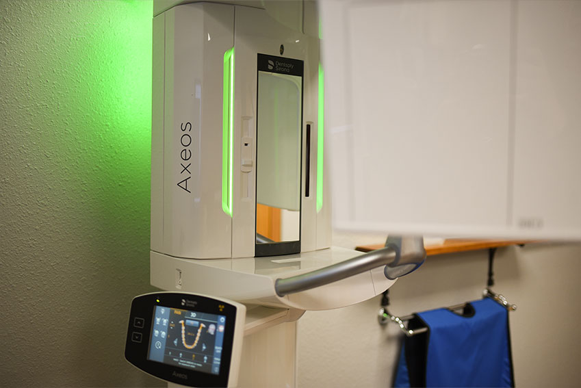
At Turman Dental, we use the Dentsply Sirona Axeos, a cone beam CT (CBCT) scanner that captures your teeth, jaws, and surrounding structures in three dimensions. Dr. Cris Turman completed CBCT training with SPEAR Education in Scottsdale, Arizona, so the images are read by someone who knows what to look for and how to apply it to your treatment plan.
- Dentsply Sirona Axeos cone beam CT
- 3D views of bone, nerves, sinuses, and airway
- SPEAR Education CBCT training
- In-office imaging, no referral needed
- Results reviewed with you on screen
What a Cone Beam CT Scan Shows
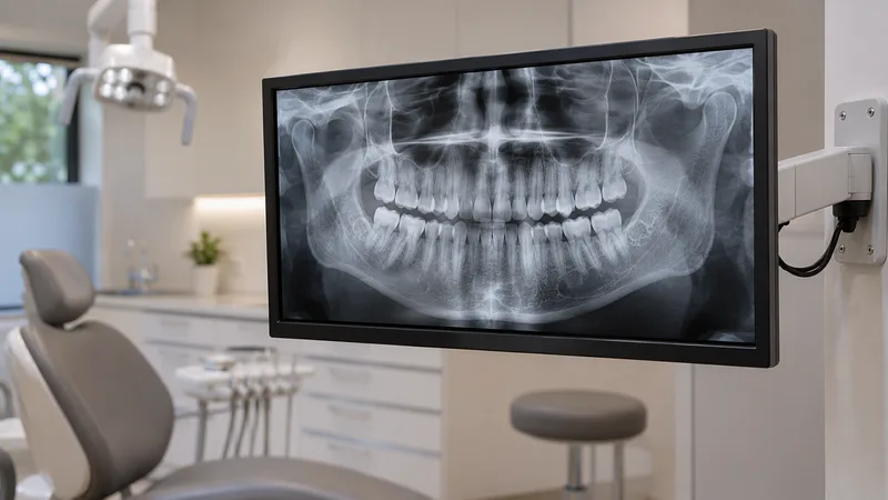
A standard x-ray flattens everything into one picture. A cone beam CT scan builds a 3D model that we can rotate, slice, and measure on screen. That helps us see:
- The amount, density, and shape of your jawbone
- The position of nerves and blood vessels
- Sinus proximity to upper tooth roots
- The shape and curvature of tooth roots
- The jaw joint (TMJ) and surrounding structures
- The airway space behind the tongue
This level of detail is especially important for procedures where millimeters matter, like placing a dental implant or planning a complex extraction.

When We Recommend 3D Imaging
Implant Planning
Good implant placement depends on knowing exactly how much bone you have and where nearby structures sit. The Axeos shows bone width, height, and density in three dimensions, so Dr. Turman can plan the angle and depth before treatment starts.
Complex Extractions
When a tooth has unusual root anatomy, sits close to a nerve, or is impacted, a 3D scan reveals the relationship between the tooth and surrounding structures. That reduces surprises during the procedure.
Endodontic Assessment
A cracked tooth or persistent infection may not show clearly on a flat x-ray. CBCT can reveal fracture lines, extra canals, and the extent of infection around the root.
TMJ and Airway Evaluation
A 3D view of the jaw joint can help us look for structural clues when you have jaw pain or clicking. Airway imaging can also be part of evaluating sleep-related breathing concerns.

Safety, Radiation, and When It Is Worth It
CBCT uses radiation, so we use it only when the diagnostic benefit justifies the exposure. For routine check-ups and cavity detection, our digital x-rays are all you need. We reserve 3D imaging for cases where the added detail will genuinely change how we plan or perform your treatment.
Please tell us if you are or might be pregnant before any imaging. We can often request copies of recent scans from another office instead of repeating them.
The scan itself takes about 15 to 30 seconds. You stand still while the scanner rotates around your head. There is no impression material and nothing to bite down on. Afterward, Dr. Turman reviews the images with you on screen so you can see exactly what we see.
Why 3D Imaging at Turman Dental
SPEAR-Trained Interpretation
Dr. Turman completed advanced CBCT training at SPEAR Education, so your images are read by someone who knows what to look for.
In-Office, No Referral
The Axeos is right here in our Linton office. No driving to a hospital or imaging center for a scan.
Fewer Trips for Rural Patients
Patients from Strasburg, Napoleon, Wishek, Hazelton, Hague, Braddock, Zeeland, and northern SD get advanced imaging without a long drive.
What to Expect During a 3D Scan
A quick, comfortable process from positioning to review
Positioning
We position you in the scanner and make sure you are comfortable. You will stand or sit depending on the area being imaged.
The Scan
The scanner rotates around your head in about 15 to 30 seconds. Hold still and breathe normally.
Image Processing
The software builds a 3D model from the scan data. It is ready for review within minutes.
Review with Your Dentist
Dr. Turman walks you through the images on screen, pointing out the structures that matter for your treatment plan.
Treatment Planning
The 3D data feeds directly into implant planning, surgical guides, and other digital workflows so your care is precise from the start.
3D Dental Scanner FAQ
Common questions about CBCT imaging at Turman Dental in Linton