


See What the Eye Cannot
The most important dental problems are often invisible on the surface. Cavities forming between teeth, bone loss creeping beneath the gumline, an infection at the tip of a root. These are the problems that x-rays reveal, and finding them early usually means simpler, less expensive treatment.
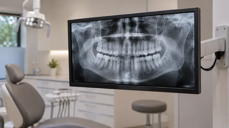
At Turman Dental, we use digital x-rays instead of traditional film. A small sensor captures the image, which appears on screen within seconds. Digital sensors typically use less radiation than film, and the images can be enlarged, contrast-adjusted, and compared side by side with previous visits.
- Lower radiation than traditional film
- Images on screen within seconds
- Enlarge, adjust, and compare images
- Easy to share with specialists
- No chemical processing or waste

What Digital X-Rays Help Us Find
A visual exam only shows the surface. X-rays show what is happening between and beneath your teeth:
- Cavities between teeth and under existing fillings
- Bone loss around teeth that can signal gum disease
- Infection at the root of a tooth
- Teeth that are impacted or not coming in properly
- The condition of existing crowns, bridges, and implants
That information shapes your treatment plan, whether you need a filling, a closer look at your gum health, or simply a reassuring result that everything looks stable.
Digital X-Rays vs. Film
Digital x-rays capture the same kind of information as film, but with real advantages for you:
- Lower radiation: Digital sensors generally need a smaller dose than traditional film to produce a usable image.
- Faster results: The image appears on screen right away, so we can review it with you during your visit instead of waiting for film to develop.
- Clearer detail: We can enlarge an area, adjust contrast, and spot subtle changes that might be missed on a small film.
- Easy to compare: Digital images from different visits can be placed side by side to track changes over time.
- Easy to share: If you need to see a specialist, images can be sent digitally instead of mailed.
- Environmentally friendlier: No developing chemicals to handle or dispose of.

Part of a Complete Imaging Toolkit
Most routine visits use standard digital x-rays, and that is all many patients ever need. For more complex cases, such as implant planning, extraction assessment, or evaluating the jaw joint, we can also take a cone beam CT scan with our Dentsply Sirona Axeos scanner for a full three-dimensional view.
Dr. Turman decides which imaging is right for your situation based on what the exam shows and what treatment is being planned. The goal is the right images at the right time, not extra images for their own sake.
Digital x-rays are most commonly taken during your dental cleaning and exam visits. They are also part of planning for deep cleanings and monitoring periodontal maintenance.
Why Patients Trust Our Digital Imaging
Instant On-Screen Results
Images appear within seconds so we can review them with you during the same visit, not at a follow-up.
Complete Imaging Options
Digital x-rays for routine care, plus a 3D CBCT scanner for complex cases. Everything is in one office.
Local Diagnostic Care
Patients from Strasburg, Napoleon, Wishek, Hazelton, Hague, Braddock, Zeeland, and northern SD get advanced imaging without a long drive.
What to Expect During Digital X-Rays
A quick, routine part of your dental visit
Sensor Placement
A small digital sensor is positioned inside your mouth. It is slightly larger than a film packet but captures data electronically.
Image Capture
The x-ray is taken in a fraction of a second. You may need a few images depending on what we are looking for.
On-Screen Review
Dr. Turman reviews the images on a monitor, pointing out what each image shows and answering your questions.
Comparison and Planning
We compare current images with previous ones to track changes, then discuss any findings and next steps.
Digital X-Rays FAQ
Common questions about dental imaging at Turman Dental in Linton